The cardiorespiratory system is in fact a transport system which enables the intake and transfer of oxygen and other substances into tissues and which eliminates the carbon dioxide and other dangerous substances from the organism. It is composed of the respiratory and circulatory system. The latter forms the subject matter of this chapter.
Organization of the circulatory system, the heart
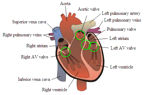
The circulatory system is made up of the heart and vessels. The heart (Fg. 17) is the central organ which works as a pump transporting oxygen throughout the blood vessels.
Figure 17 Structure of the heart
The heart activity
Like the skeletal muscles the heart is controlled by electrical impulses generated by heart.
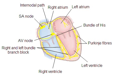
The conduction system of the heart is formed by the sinus (sinoatrial) node generating the fastest electrical impulses. Under normal circumstances the heart is controlled by the sinus node, a natural pacemaker which under resting conditions generates regular impulses within the range of 60-100*min-1. Electrical impulses then further spread into the atrioventricular node, bundle of Hiss, Tawara's nodes and Purkinje fibres (Fig. 18).
Figure 18 Conduction system of the heart
Physiological parameters of the circulatory system
Heart rate (HR) = a number of contractions per minute, given in beats per minute.
Stroke volume (Qs) = volume of blood pumped from the heart through one systole, given in ml.
Cardiac output (Q) = the volume of blood pumped from the heart during one minute, given in 1/min.
Blood pressure (BP) = is the pressure exerted by circulating blood upon the walls of blood vessels. During each heartbeat, BP varies between a maximum (systolic) and a minimum (diastolic) pressure; given in mmHg.
Reaction and adaptation of the circulatory system to workload
Heart rate
The value of resting heart rate tends to be about 70 beats/min in the average middle-aged population. HR decreases in proportion with rising age.
Response: Heart rate increases with workload (Fig. 19). Under constant workload HR rises dramatically and then it stabilizes. Under workload of increasing intensity, HR increases proportionally. If an individual carries out a movement activity of maximum intensity, the HRmax is reached. The HRmax value of a 20-year old man is about 200 beats per min. In the absence of workload, the HR returns to the original values.
Figure 19 Graph showing various HR values under workload
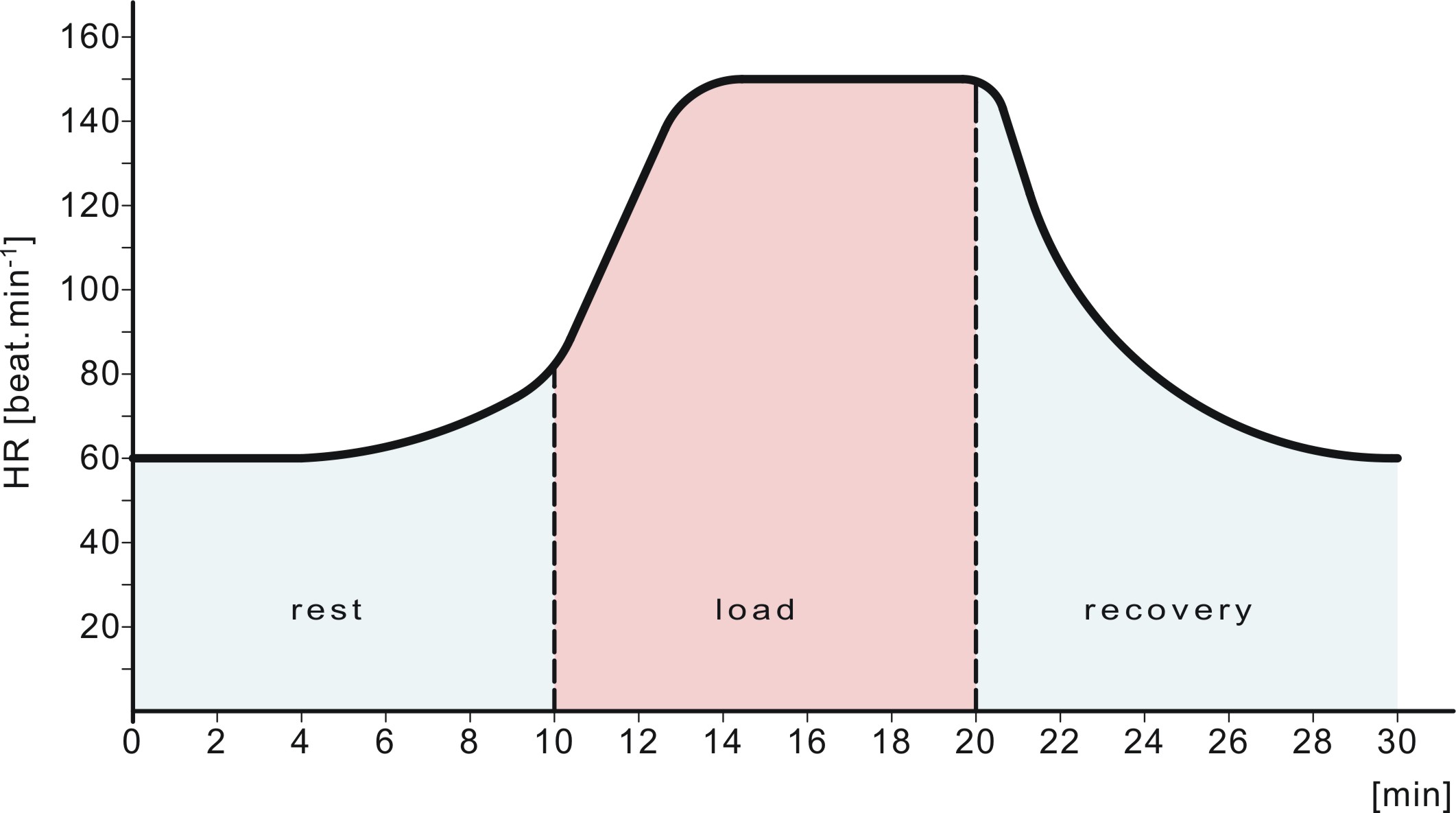
The HR values may be estimated based on the formula of 220-age.
Adaptation: The endurance trained individuals show the so called vagotonia – reduction in the resting HR values to the region under 50 beats/min. For example, elite long-distance runners, cyclists, cross country skiers, etc. show values of around 30 beats/min. This is given by the heart hypertrophy (explained later).
Heart rate may be detected using the following methods:
- palpation on the radial artery
- listening at the apex of the heart
- through electrical apparatuses
HR is best detected through palpations on the radial artery. We use 2-3 fingers to feel for the artery. As a thumb tends to contain a large artery itself, we do not use it to palpate the artery. Heart rate is measured for 20 seconds, the value obtained is multiplied by 3.
We assess the heart rate through listening most frequently at the apex of the heart using sthetoscope (normally at the doctor's).
The most frequently used apparatuses to measure HR are the so called sport-testers. There are many kinds of them, of which the latest ones include built-in GPS. In addition to measuring HR, they also measure the speed and distance of running, etc. A sport-tester consists of a chest belt with two electrodes which read the electrical activity of the heart in a similar way to ECG. The values measured show on a wrist watch. The values may be downloaded to a computer and thus it is possible to record and evaluate the training practice.
The HRV apparatus to assess the heart rate variability works similarly to a sport-tester. Like the latest sport-testers it records the value for each heart beat.
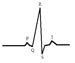
ECG reads the electrical activity of the heart using electrodes. The most frequently used one in the modern medicine is the 12-lead ECG where the curve consists of waves and swings of a typical shape and duration. Here we distinguish the P-wave (atrial depolarization), complex QRS (ventricular depolarization) and the T-wave (ventricular repolarization) – Fig. 20. The HR may easily be calculated; the interval between two adjacent R-swings equals the distance of two adjacent beats.
Figure 20 Generation of excitation and the ECG curve
Stroke volume
The resting values of the systolic volume in the average man are in the region of 70 ml.
Response: Under workload the values tend to rise, depending again on the workload intensity. The maximum values may be around 130 ml.
Adaptation: In endurance trained individuals the resting values are in the region of 100 ml. This increase results from excentric heart hypertrophy during which heart ventricles are enlarged and the heart is able to expel more blood at the same time. This makes it possible for the person to achieve higher values of up to 200 ml under workload.
Cardiac output
The resting values of the minute heart volume are about 5 litres in men and a little less in women.
Response: The values grow in proportion with the rising intensity of workload. Under maximum intensity the values exceeding 25l/min may be detected.
Adaptation: In endurance trained individuals the values increase up to the level of 35l/min. This again is given by the heart hypertrophy.
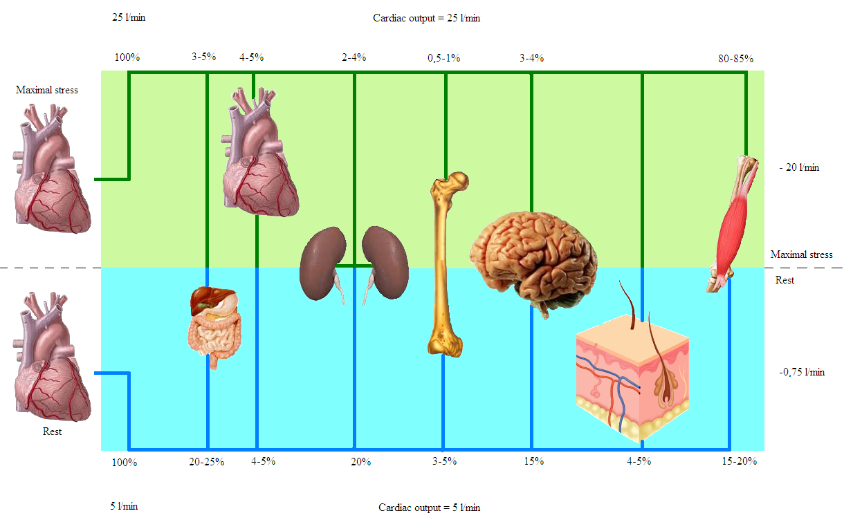
The following picture (Fig. 21) shows blood redistribution under resting conditions and high intensity workload.
Figure 21 Blood distribution in organs under resting conditions and during heavy exercise
Blood pressure
The ideal blood pressure values are in the region of 120/80 mmHg. Hypertension, i.e. increased blood pressure, occurs if the pressure exceeds the level of 140/90 mmHg while hypotension, decreased blood pressure is that of 100/65 mmHg and lower.
Response: The blood pressure value changes under workload depending on its type and intensity. Under dynamic workload conditions the systolic pressure increases. Maximum values may be detected under submaximal workload intensity where they could exceed the 200 mmHg level. The diastolic pressure, however, may decrease under submaximal intensity. The static workload normally results in the increase of both the systolic and diastolic pressure.
While in the past classic mercury based apparatuses were used, nowadays the blood pressure may be measured through automatic or semiautomatic tonometres which are attached to an arm, wrist or finger. To detect the value of pressure a sthetoscope is used.